Sunday Poster Session
Category: Colon

Abel Sanchez, MD, MSc
Hospital Roosevelt / Gastri-k
Guatemala City, San Marcos, Guatemala
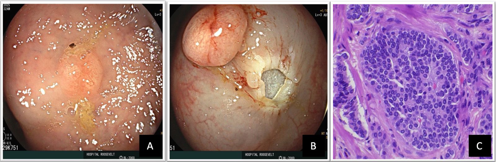
Rectal neuroendocrine tumors are observed as small round polypoid lesions, characterized by a smooth mucosa of normal appearance, the atypical findings are semi-pedunculated appearance, hyperemia, central depression, erosion and ulceration. They are found between 4 and 10 cm above the dentate line, on the front or lateral wall of the middle rectum, located in the submucosa, without invading the muscularis propria. The risk of metastasis ranges between 3-60%, therefore, early detection and elimination is important.
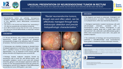
The diagnosis was established by histology and immunohistochemistry results and classified according to the proliferation index (mitotic count and Ki67-related proliferation index). Treatment will depend on whether it is localized, locally advanced or advanced metastatic disease