Sunday Poster Session
Category: Colon

Charles B. Woodall, DO
Louisiana State University Health Sciences Center New Orleans
New Orleans, LA
Rectal leiomyomas and neuroendocrine tumors (NET) are both considered rare with incidences of 0.05% and 0.17%, respectively. When present, both are typically discovered incidentally during routine endoscopy but can cause symptoms and complications such as metastasis, obstruction, hemorrhage, and perforation if not discovered early and allowed time to advance. The simultaneous existence of a leiomyoma and NET in the rectum has not previously been described in the literature to date and is an exceedingly rare occurrence.
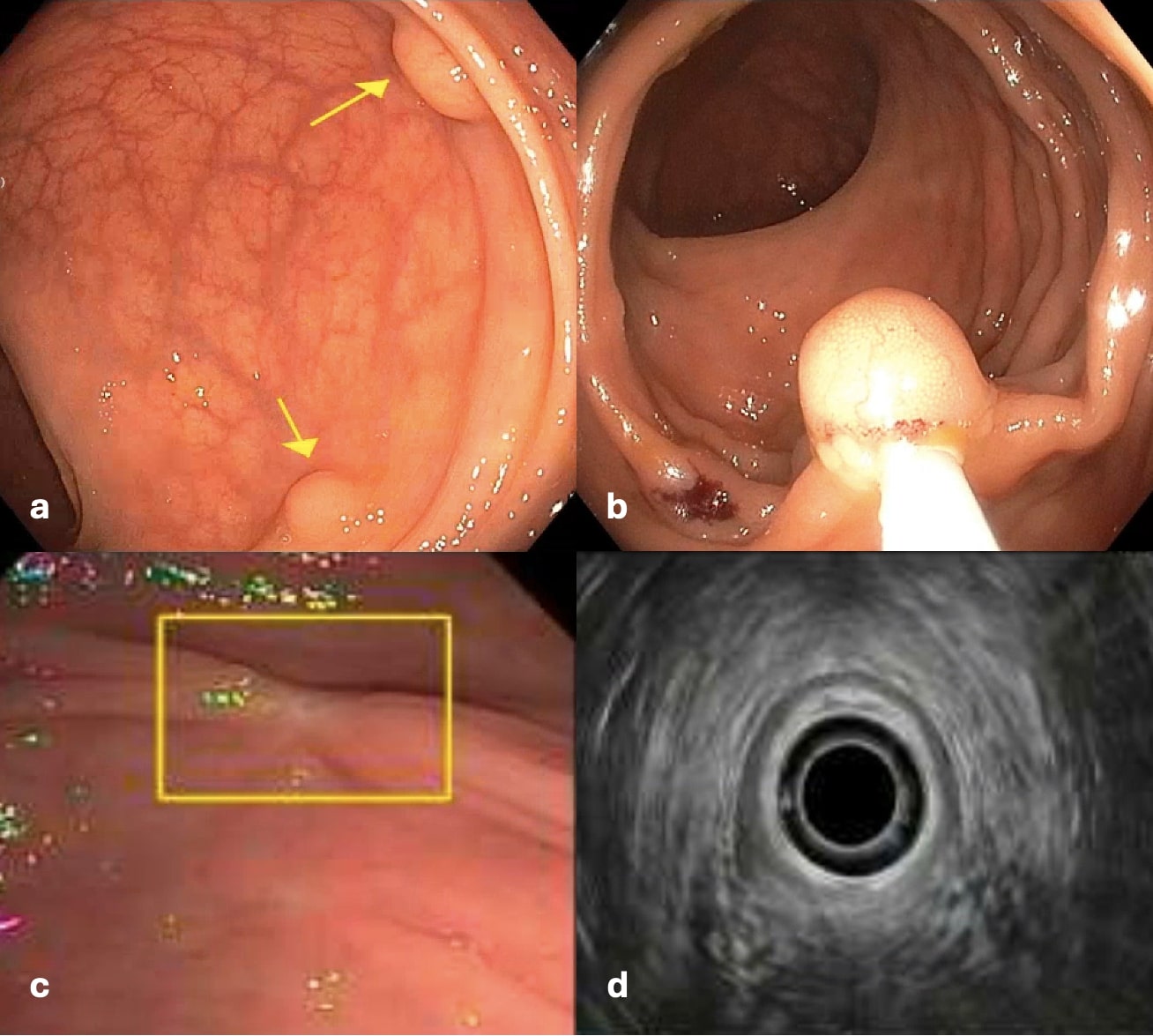
Our patient is a 52-year-old female with a history of type II diabetes mellitus, hypertension, and obesity who was found to have two rectal polyps during index screening colonoscopy. The 6 mm lesion was removed via cold snare, and the 8 mm lesion with submucosal appearance was removed via hot snare. Pathology identified the lesions as a leiomyoma and a grade I NET, respectively. She subsequently underwent EUS with repeat biopsy that was negative for any residual or recurrent NET as well as any lymphatic involvement. Repeat lower EUS for surveillance was planned for a year from that time.
Rectal leiomyomas and rectal NETs are both rare tumors, individually, making their concurrent existence an exceptionally rare phenomenon. While leiomyomas in general are more common in females, rectal leiomyomas occur more commonly in males with one study demonstrating a 2.5:1 ratio. Incidental diagnosis via routine endoscopy is involved in most cases of both, but symptomatic presentations can be seen with more advanced lesions. Differentiation of leiomyomas from leiomyosarcomas and gastrointestinal stromal tumors is essential due to significant prognostic differences. Treatment via surgical resection is indicated for tumors involving the submucosa. Leiomyomas originating from the muscularis, however, can be resected via endoscopy. Endoscopic resection can be achieved utilizing EMR to better differentiate mucosal layers and margins for removal with electrosurgical (hot) snare, or a bite-on-bite technique utilizing jumbo forceps can be employed. While preoperative assessment of rectal NETs can be more complex due to the potential for metastasis, management is similar to that of rectal leiomyomas. EUS is often performed to help classify lesions and formulate a treatment plan prior to intervention and following endoscopic resection to assess for eradication.