Tuesday Poster Session
Category: Colon


Hassam Ali, MD
East Carolina Gastroenterology
Greenville, NC
We conducted a retrospective analysis of polyp-level histopathologic data from screening colonoscopies performed between 2019–2023. Eligible patients were average-risk individuals aged ≥45 years undergoing colonoscopy for colorectal cancer screening. All polyps were resected and categorized by size: 1–5 mm, 6–9 mm, 10–14 mm, 15–20 mm, and >20 mm. Chi-square test was used to assess any statistical differences.
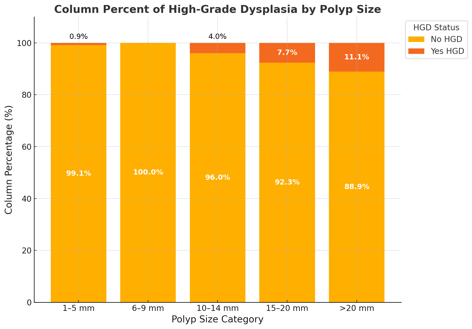
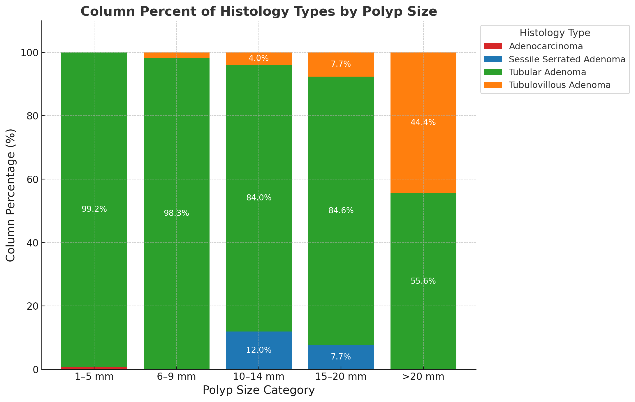
A total of 222 polyps were identified from average-risk screening colonoscopies. Overall, 4 polyps (1.8%) contained HGD, including 1 of 117 polyps (0.85%) in the 1–5 mm group, 1 of 25 (4.0%) in the 10–14 mm group, 1 of 13 (7.7%) in the 15–20 mm group, and 1 of 9 (11.1%) in the >20 mm group (p = 0.05) (Figure 1). No HGD was found in the 6–9 mm size group. One invasive adenocarcinoma (0.45%) was detected in a 1–5 mm polyp; all other size categories had zero cases of cancer (p = 0.92). Tubular adenomas were the most common subtype, accounting for 210 of 222 polyps (94.6%), including 99.2% of 1–5 mm polyps and 98.3% of 6–9 mm polyps. Tubulovillous adenomas made up 44.4% of >20 mm polyps, compared to just 1.7% of 6–9 mm and 4.0% of 10–14 mm polyps. Sessile serrated adenomas were identified in 12.0% of 10–14 mm and 7.7% of 15–20 mm polyps, but were absent in polyps < 10 mm or >20 mm (Figure 2). HGD was more frequent in men (75%) and Black patients (50%) despite comprising 62.6% and 39.2% of the cohort, respectively. All HGD lesions were right-sided, located in the ascending colon (75%) or cecum (25%), suggesting possible demographic and anatomic variation.
In this average-risk screening cohort, high-grade dysplasia was detected in nearly 2% of polyps, including diminutive lesions, with one 5 mm polyp showing invasive cancer. These findings challenge assumptions of minimal risk in small polyps in the United States population.