Monday Poster Session
Category: Colon

Matthew A. Kalloo, MD
Harlem Hospital Center
New York, NY
Müllerian tissues originate from paramesonephric ducts and differentiate into uterus, fallopian tubes, cervix, and upper vagina. Müllerian adenocarcinomas account for 2% of gynecologic neoplasms. GI metastases usually occurs to the rectosigmoid or rectovaginal septum. We report a case of Mullerian adenocarcinoma presenting as a large colonic mass.
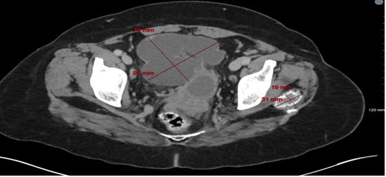
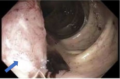
A 59-year-old female presented with 2 weeks of fatigue and abdominal pain. Her last menstrual period was 13 days ago. She was afebrile, BP 153/75, pulse 138. She was pale and lethargic with a benign nondistended abdomen. Hb was 4.6, MCV 68.8, WBC 9.9k, and platelets 498k. Ferritin was 18 ng/ml, iron 11 μg/dl, iron saturation 3%, Retics 3.28% and LDH 283 U/L. CT showed multiple cystic heterogeneous irregular abdominopelvic masses; in the area of the ovaries, adjacent to the uterus, LUQ, RUQ, and epigastric, most 5-10 cm, with multiple scattered pelvic, mesenteric, and inguinal lymph nodes. (Fig.1). She was admitted to ICU where she was transfused. EGD was normal. Colonoscopy showed a fungating, non-obstructive, non-bleeding, partially circumferential mass in the ascending colon, 20 cm in length (Fig. 2). Biopsy revealed a poorly differentiated adenocarcinoma of primary Müllerian origin. CA-125 was elevated at 2006 U/ml (< 38); CA 19-9 and CEA were both normal. She declined additional work up. She was discharged with Hb of 8.1. She presented 2 months later to an outside facility with multifocal small and large bowel obstruction due to peritoneal carcinomatosis. She was again offered surgery and chemotherapy but declined.