Carla Barberan Parraga, MD1, Syed Mujtaba Baqir, MD1, Tanuj Chokshi, DO1, Neha Sharma, MD1, GianPaolo Piccione, DO2, Nicole Villa, MD3, Samar Pal S. Sandhu, MBBS1, Manol Jovani, MD, MPH4 1Maimonides Medical Center, Brooklyn, NY; 2Maimonides Medical Center - Open School UNITED STATES - Open School, Staten Island, NY; 3NYC Health + Hospitals/Metropolitan, New York, NY; 4GastroHealth, Florida International University, Miami, FL
Introduction: Subepithelial lesions (SEL) have become more prevalent findings with increased adherence to colorectal cancer screening through colonoscopy. SEL can be present throughout the gastrointestinal tract and can represent benign or malignant findings. Several studies have described their malignant potential and their correlation with endoscopic features, as well as proposed techniques for accurate characterization, including the use of endoscopic ultrasound and fine-needle biopsy. We describe a case of a subepithelial lesion removed by endoscopic full-thickness resection with endoscopic findings suggestive of a granulomatous reaction.
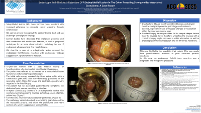
Case Description/Methods: A 47-year-old female patient with a past medical history of hypothyroidism who immigrated from Russia 30 years ago. The patient was referred for a subepithelial lesion resection found in an initial screening colonoscopy. The initial colonoscopy showed significant active colitis with a submucosal granuloma and a non-necrotizing granuloma in the ascending colon. Stains for fungal and acid-fast organisms were negative in the initial biopsy. The patient denied any abdominal pain, nausea, vomiting, or diarrhea. No other gastrointestinal symptoms, unintentional weight loss, or history of parasitic infections. A repeat colonoscopy showed a 1 cm subepithelial nodule with overlying normal-appearing mucosa with a non-attached yellow hue. A full-thickness resection was successfully performed, and the tissue was sent for evaluation. The pathology report described a necrotizing granuloma within the muscularis propria. The granuloma shows sections of a worm suggestive of Strongyloides.
Discussion: Small colonic SEL are mostly considered benign, and despite their low malignant potential, pathology confirmation is essential, especially in the case of mucosal changes or localization within the muscular mucosa layer. Standard biopsy techniques fail to sample deeper lesions than the mucosal layer thus, unroofing the mucosa with posterior biopsy might represent a viable technique as well as endoscopic submucosal resection and full thickness resections.
Figure: Figure 1. Subepithelial nodule with overlying normal-appearing mucosa with a non-attached yellow hue.
Disclosures:
Carla Barberan Parraga indicated no relevant financial relationships.
Syed Mujtaba Baqir indicated no relevant financial relationships.
Tanuj Chokshi indicated no relevant financial relationships.
Neha Sharma indicated no relevant financial relationships.
GianPaolo Piccione indicated no relevant financial relationships.
Nicole Villa indicated no relevant financial relationships.
Samar Pal Sandhu indicated no relevant financial relationships.
Manol Jovani: Pentax Medical – Consultant.
Carla Barberan Parraga, MD1, Syed Mujtaba Baqir, MD1, Tanuj Chokshi, DO1, Neha Sharma, MD1, GianPaolo Piccione, DO2, Nicole Villa, MD3, Samar Pal S. Sandhu, MBBS1, Manol Jovani, MD, MPH4. P3586 - Endoscopic Full-Thickness Resection of a Subepithelial Lesion in the Colon Revealing <i>Strongyloides</i>-Associated Granuloma, ACG 2025 Annual Scientific Meeting Abstracts. Phoenix, AZ: American College of Gastroenterology.