Tuesday Poster Session
Category: Colon

Ruchir Paladiya, MBBS
University of Connecticut School of Medicine
Farmington, CT
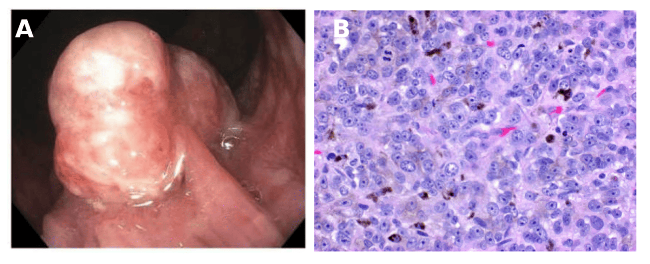
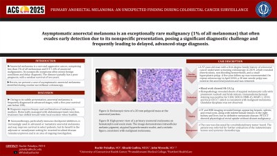
A 57-year-old man with a first-degree family history of colorectal cancer underwent screening colonoscopy in 2019, which revealed diverticulosis, non-bleeding hemorrhoids, and a small hyperplastic polyp. A five-year follow-up was recommended. On repeat colonoscopy in April 2024, a 20 mm sessile polyp was noted at the anorectal junction and was removed. Histopathology revealed sheets of atypical melanocytic cells with prominent nucleoli and brisk mitosis. Immunohistochemical staining was positive for S100, SOX10, HMB-45, MART-1, CD10, and focal CD117, which is consistent with malignant melanoma. Glandular dysplasia was not observed.
CT and MRI imaging revealed benign-appearing hepatic, splenic, and renal cysts, along with indeterminate lesions in the right kidney and liver, but no definitive metastatic disease. PET/CT showed physiological rectal uptake without distant malignancy. Physical examination results were unremarkable. The case was discussed by a multidisciplinary tumor board. The patient was referred for further evaluation of the indeterminate lesions and systemic chemotherapy.