Shantanu Anant Shahane, MD1, Rajkumar P. Wadhwa, MD1, Aathira Ravindranath, MD1, Kushan Sengupta, MD1, Raghuram Naik, MD1, Rakesh JP, MD1, Smruti Sangam, MD1, Madhuri R. Wadhwa, MD2 1Apollo BGS Hospitals, Mysore, Karnataka, India; 2Accura Diagnostics and Healthcare, Mysore, Karnataka, India
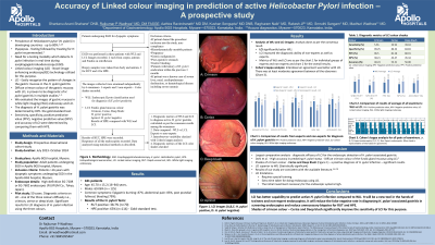
Introduction: Prevalence of Helicobacter pylori (H.pylori) infection worldwide varies from 50 to 80%. Test and treat strategy for H. pylori have been recommended for all patients with symptoms. Linked color imaging (LCI) is a novel Image enhanced endoscopy (IEE) technology which has been utilized for this purpose. In this study we tested the accuracy of LCI in our center in predicting H. pylori infection.
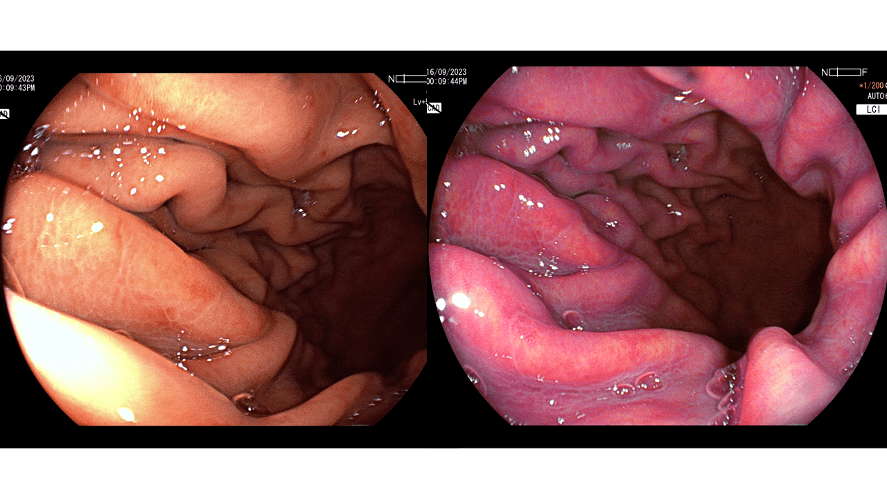
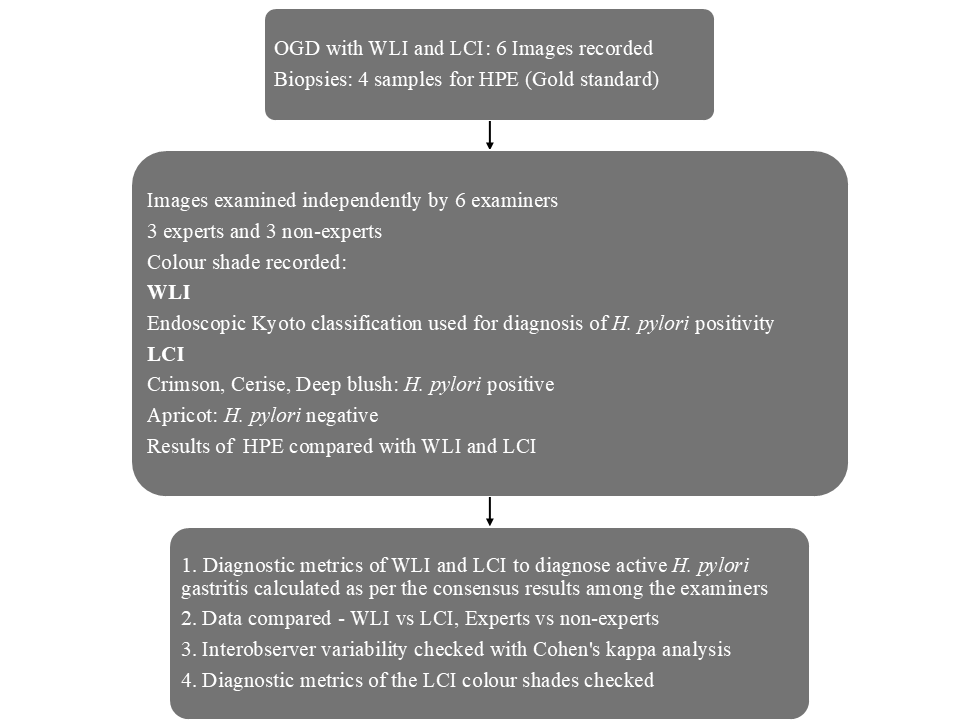
Methods: Study Design: Prospective observational cohort study. Inclusion Criteria: Patients more than 18 years old undergoing esophagogastroduodenoscopy for dyspeptic symptoms in whom H.pylori infection was suspected. Study protocol: Pilot study was conducted among experts with images of 50 patients in white light imaging endoscopy (WLI) and LCI (Figure 1). The ability to detect H. pylori positivity on LCI improved when shades of crimson namely cerise and deep blush were included. Final study protocol was followed as per figure 2.
Results: Study involved 181 patients (69% males) aged 45.7 years (16-84). Epigastric burning was the most common symptom followed by abdominal pain. Histopathological examination (HPE) was positive for H.pylori gastritis in 114(63%) patients. The sensitivity, specificity, PPV, NPV, accuracy of WLI for diagnosing H.pylori infection are 70.27%, 47.34%, 64.03%, 54.17%, 60.4% respectively, and that of LCI are 83.22%, 71.24%, 82.89% 71.64%, 78.73% respectively (LCI better than WLI. (P< 0.001)). Experts performed better than non-experts. Experts and non-experts performed better with LCI than WLI (P< 0.001). For WLI images, moderate agreement was noted amongst the experts and moderate to fair between the experts and the non-experts and amongst the non-experts. Interobserver agreement was better with LCI. There was at least moderate agreement between all the observers. All three shades of crimson had specificity of more than 84% for diagnosing H.pylori infection but the sensitivity of individual colors is poor. LCI has a good performance in all the diagnostic metrics when data of all three shades of crimson were combined.
Discussion: LCI can be used to predict the diagnosis of H. pylori gastritis with an accuracy of 79%. When the color palette of crimson is extended to include cerise and deep blush shades performance of LCI increases compared to crimson alone.
Figure: WLI and LCI images - H.pylori positive
Figure: Final study protocol
Disclosures:
Shantanu Anant Shahane indicated no relevant financial relationships.
Rajkumar Wadhwa indicated no relevant financial relationships.
Aathira Ravindranath indicated no relevant financial relationships.
Kushan Sengupta indicated no relevant financial relationships.
Raghuram Naik indicated no relevant financial relationships.
Rakesh JP indicated no relevant financial relationships.
Smruti Sangam indicated no relevant financial relationships.
Madhuri Wadhwa indicated no relevant financial relationships.
Shantanu Anant Shahane, MD1, Rajkumar P. Wadhwa, MD1, Aathira Ravindranath, MD1, Kushan Sengupta, MD1, Raghuram Naik, MD1, Rakesh JP, MD1, Smruti Sangam, MD1, Madhuri R. Wadhwa, MD2. P5144 - Accuracy of Linked Color Imaging (LCI) in Prediction of Active <i>Helicobacter pylori</i> Infection - A Prospective Study, ACG 2025 Annual Scientific Meeting Abstracts. Phoenix, AZ: American College of Gastroenterology.