
Tuesday Poster Session
Category: Colon
Jasneet Gill, MBBS, MD
North Knoxville Medical Center
Knoxville, TN
Colorectal cancer (CRC) is one of the most common malignancies in the U.S., typically presenting as a single primary tumor. However, the occurrence of dual primary colonic malignancies is a rare phenomenon. The reported frequency of synchronous Colorectal cancer is around 3.5% of all CRC and mean age is the seventh decade.
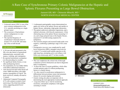
A 38-year-old male presented to the ER with a two-day history of generalized abdominal pain, associated with nausea, vomiting, and constipation. He reported intermittent constipation over the preceding two months. No significant family history, or any other predisposing factors. On examination, he was was mildly febrile with diffuse abdominal tenderness. Labs showed elevated white blood cell count of 14000 microliters with slight anemia, hemoglobin of 9.8g/dl . His lactic acid level, initially elevated at 2.1mmol. Abdominal Computed Tomography (CT) with contrast revealed a focal narrowing of the distal descending colon, suggesting partial bowel obstruction. A subsequent gastrographic enema demonstrated an apple-core lesion at the splenic flexure and another at hepatic flexure, indicating a high-grade obstruction. The patient underwent exploratory laparotomy, and a subtotal colectomy with ileocolic anastomosis. A firm obstructing mass at the splenic flexure measuring 3.5 cm, and a large polypoid mass at the hepatic flexure, measured 4.5 cm both raising suspicion for malignancy. The resection margins and resected lymph nodes were negative, confirming a pathologic stage of IIA colon cancer. Postoperative recovery was complicated by small bowel obstruction (SBO), managed conservatively. A chest CT revealed small nodular opacities in the right lung base, prompting active surveillance with follow-up colonoscopy and CT in three months.