Monday Poster Session
Category: Colon
.jpg "Courtney O'Donnell, BS, MBA photo")
Courtney O'Donnell, BS, MBA
Marian University College of Osteopathic Medicine
Indianapolis, IN
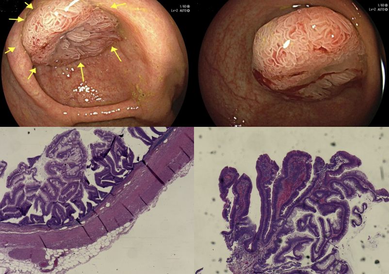
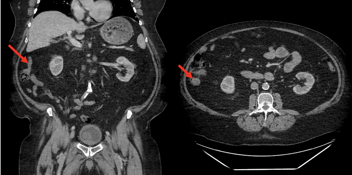
A 72-year-old asymptomatic male underwent routine surveillance colonoscopy. Endoscopy revealed a polypoid, villous-appearing lesion at the appendiceal orifice. Initial biopsy pathology demonstrated a villous adenoma. However, due to the endoscopic appearance of an appendiceal lesion, a contrast-enhanced CT scan was obtained. Imaging showed a dome-shaped protrusion at the base of the cecum consistent with the volcano sign. The patient underwent appendectomy. Gross pathology revealed a dilated appendix, and final histology confirmed a LAMN with negative margins. Notably, the lesion exhibited prominent villous mucosal architecture corresponding to the endoscopic appearance - distinct from the mucin-filled morphology typically associated with the volcano sign.
This case illustrates a rare and distinctive endoscopic presentation of the volcano sign, characterized by a villous lesion at the appendiceal orifice rather than the classic mucin-filled dome. The unusual villous appearance was initially diagnosed as a villous adenoma. Further imaging and surgical pathology identified a LAMN - a typically slow-growing lesion with low malignant potential -emphasizing the need for early recognition to guide management and prevent complications like mucin spread. This case suggests that villous morphology at the appendiceal orifice should raise suspicion for the volcano sign and underlying mucinous neoplasm, even in asymptomatic patients. Awareness of this unique feature may improve early diagnosis and expand understanding of LAMN presentations.