Monday Poster Session
Category: Colon
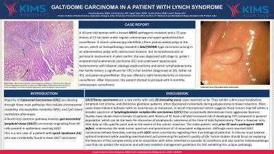

Vishal Pande, MBBS
Krishna Institute of Medical Sciences
Hyderabad, Telangana, India
Majority of Colorectal Carcinomas (CRC) can develop through three main pathways that include chromosomal instability, microsatellite instability (MSI), and CpG island methylator phenotype. A third less common pathway involves gut-associated lymphoid tissue (GALT) carcinomas originating from M-cells present in epithelium covering GALT. This is a rare case of a patient with Lynch Syndrome (LS) who was incidentally found to have GALT carcinoma.
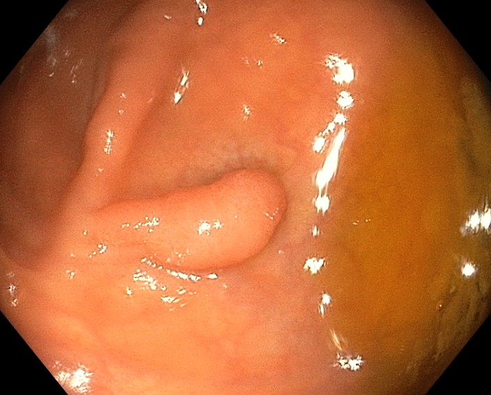
A 43-year-old woman with a known MLH1 pathogenic mutation and a 15-year history of LS has been under regular colonoscopic and upper gastrointestinal surveillance. A recent colonoscopy identified a 9mm pedunculated polyp in the cecum, which on histopathology revealed a GALT/DOME-type carcinoma arising in an adenomatous polyp with submucosal invasion, but no lymphovascular or perineural involvement. 4 years earlier, she was diagnosed with stage Ia, grade I endometrioid endometrial carcinoma (EC) and underwent laparoscopic hysterectomy with bilateral salpingo-oophorectomy and pelvic lymphadenectomy. Her family history is significant for CRC in her brother (diagnosed at 30), father (at 45), and paternal grandfather. She was offered a right hemicolectomy or intensive surveillance. After discussion, the patient elected to proceed with 6-monthly colonoscopic surveillance.
GALT tumors are characterized by submucosal localization, prominent lymphoid infiltrate, and dilated glands lined by columnar epithelium. Initially described as having dome-like appearance GALT carcinomas can present various morphologies, including polypoid (this case) and plaque-like lesions and are often associated with pre-existing conditions such as Ulcerative colitis, LS, or Familial Adenomatous Polyposis. A literature review done in 2019 documented 23 cases and reviewed 20 patients, 11 patients found to have GALT carcinoma at surveillance colonoscopy biopsy and 9 patients at diagnostic colonoscopic biopsy. GALT carcinomas are rare because GALT domains occupying colorectal mucosal areas are minute. The patient discussed in this case report had a 15 years history of LS, associated with CRC and EC. LS is associated with MSI and mismatch repair deficiency, while GALT carcinomas are typically microsatellite stable. GALT carcinomas have excellent prognosis as no cases have been reported to have metastasis. Following the latest available guidelines and given the good prognosis, our patient was managed with polypectomy and was advised for long term follow up.