John Michael Vincent Coralde, MD1, Alyssa Vergara, MD2, Ryan Burd, DO2, Ryan Gilbertson, MD2, Ana Soriano-Munoz, MD2, Hannah SoCal MEC. Haughn, DO2, Indraneel Chakrabarty, MD2 1Southwest Healthcare MEC, Murrieta, CA; 2Southwest Healthcare MEC, Temecula, CA
Introduction: Helicobacter pylori (H. pylori) is a bacterium that commonly resides in the stomach affecting nearly half the global population. While H pylori is a well-documented risk factor for gastric malignancies, including mucosa-associated lymphoid tissue (MALT) lymphoma, most individuals with H. pylori are asymptomatic. This case highlights the diverse manifestations of H. pylori, the difficulty that it introduces to the diagnosis process, and the need for further research into this complex gastrointestinal malignancy.
Case Description/Methods: 38-year-old female with no past medical hx who presented to the emergency department with dyspnea on exertion, epigastric discomfort, early satiety, and abdominal bloating.A detailed history revealed intermittent abdominal symptoms over the past 2–3 years, which she attributed to dietary factors and for which she sought no prior treatment. Initial laboratory investigations showed elevated ESR and CRP. Tumor markers were also significantly elevated with CA 125 at 178 U/mL, CA 19-9 at 112,263 U/mL, and CEA at 88.8 ng/mL. Imaging findings showed bilateral enlarged ovaries, small-volume ascites, scattered enlarged mesenteric lymph nodes, subtle omental nodularity, and irregular thickening of the gastric wall. Endoscopic evaluation revealed a large fungating mass in the gastric body. Histopathological analysis demonstrated Helicobacter pylori infection and atypical cells concerning MALT lymphoma (MALToma). The patient was started on bismuth-based quadruple therapy (BQT) for H. pylori eradication. She was subsequently discharged with referrals for oncology follow-up for further evaluation and management.
Discussion: The case highlights an exceedingly rare association between H-pylori associated gastritis, MALToma and Krukenberg tumor, likely owing to the inherent direction of lymphatic flow. Lymphatic flow, the means by which MALToma typically spreads, moves from the body and fundus of the stomach to the pancreaticosplenic lymph nodes, through the celiac lymph nodes and ultimately draining into the thoracic duct. As such, this case provides exposure to the possibility of caudal metastasis and resistance to lymphatic flow of MALToma. Ovarian Kruckenberg tumors as a sequelae of MALToma account for less than 1% of studied metastatic sites. This case highlights the diverse manifestations of H. pylori and the need for further research into this complex gastrointestinal malignancy.
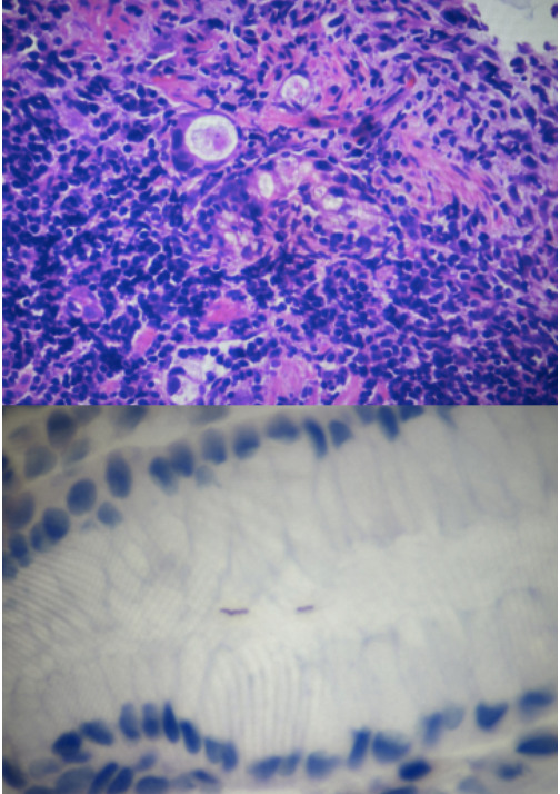
Figure: Microscopic findings seen in severe chronic H. pylori associated gastritis: with severe chronic inflammatory infiltrates within the gastric mucosa with deeper focal areas of acute inflammation, erosion, and atypical cells with large hyperchromatic nuclei and reactive glandular atypia.
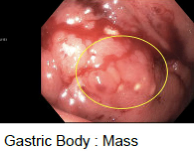
Figure: Patient’s EGD findings showing large, fungating , circumferential mass oozing with blood in the atrophic gastric body.
Disclosures:
John Michael Vincent Coralde indicated no relevant financial relationships.
Alyssa Vergara indicated no relevant financial relationships.
Ryan Burd indicated no relevant financial relationships.
Ryan Gilbertson indicated no relevant financial relationships.
Ana Soriano-Munoz indicated no relevant financial relationships.
Hannah Haughn indicated no relevant financial relationships.
Indraneel Chakrabarty indicated no relevant financial relationships.
John Michael Vincent Coralde, MD1, Alyssa Vergara, MD2, Ryan Burd, DO2, Ryan Gilbertson, MD2, Ana Soriano-Munoz, MD2, Hannah SoCal MEC. Haughn, DO2, Indraneel Chakrabarty, MD2. P1352 - Unmasking the Consequences of Chronic <i>H. pylori</i> Infection: MALT Lymphoma Presenting with Peritoneal Carcinomatosis and Krukenberg Tumor, ACG 2025 Annual Scientific Meeting Abstracts. Phoenix, AZ: American College of Gastroenterology.